Why Capnography Catches Anesthesia Problems Before Pulse Oximetry
How capnography detects hypoventilation, esophageal intubation, and circuit disconnects minutes before pulse oximetry. Plus how to secure your CO2 sensor to prevent false readings.
What Capnography Actually Tells You
Capnography measures carbon dioxide concentration in respiratory gases, breath by breath. The number everyone watches is ETCO2 (end-tidal CO2), the peak CO2 at the end of each expiration. That number tells you how well the patient is ventilating, and indirectly, how well the cardiovascular system is moving CO2 from tissues to the lungs.
Normal ETCO2 under anesthesia runs 35 to 45 mmHg for dogs. Cats tend to sit a bit lower, around 28 to 35 mmHg. Anything above 60 mmHg means the patient isn’t moving enough gas and needs intervention (manual breaths or lighter anesthetic depth). Below 20 mmHg is concerning for hyperventilation, low cardiac output, or cardiac arrest.
But the number is only half the story. The capnogram waveform provides real-time information about airway patency, breathing patterns, and circuit integrity that no other single monitor can give you.
Why SpO2 Isn’t Enough on Its Own
Most people trust the pulse oximeter. It’s familiar, it’s on every patient, and a dropping SpO2 gets everyone’s attention. The problem is that SpO2 is a lagging indicator. It only drops after the patient’s oxygen reserves are already depleted.
When ventilation stops or becomes inadequate, the lungs still have a reserve of oxygen sitting in functional residual capacity. That buffer can keep SpO2 looking perfectly normal for two to five minutes, sometimes longer if the patient was pre-oxygenated. Meanwhile, the patient isn’t actually breathing effectively.
ETCO2 changes within one breath. The moment something goes wrong with ventilation (disconnected circuit, kinked tube, respiratory depression), the capnograph shows it immediately.
A closed claims analysis of 1,097 anesthesia injuries and deaths (Tinker et al., Anesthesiology, 1989) found that pulse oximetry and capnography together could have prevented 93% of the preventable negative outcomes. SpO2 catches late-stage desaturation. Capnography catches the ventilation problem that causes the desaturation, minutes before the oxygen numbers start to slide.
The Things Only a Capnograph Will Show You
Some of the most dangerous anesthetic events are silent without capnography:
Esophageal intubation. If the tube goes in the esophagus instead of the trachea, you get a flat capnogram. No CO2 being expired from the lungs, detectable within two breaths. Without capnography, this can go unrecognized for several minutes until SpO2 finally drops.
Circuit disconnect. ETCO2 drops from normal to zero instantly. Unambiguous.
Hypoventilation. A gradually climbing ETCO2 trend means the patient isn’t moving enough gas. This can creep up slowly from excessive anesthetic depth or opioid-induced respiratory depression, easy to miss if you’re not watching the number.
Rebreathing. When the baseline CO2 never returns to zero during inspiration, the patient is breathing back in their own expired gas. Usually means exhausted soda lime or a stuck one-way valve.
Cardiac arrest. ETCO2 drops below 10 to 15 mmHg when cardiac output ceases. During CPR, it’s actually the best real-time indicator of whether your compressions are effective and whether you’re getting return of spontaneous circulation.
Sidestream vs. Mainstream: Which One and Why
Mainstream sensors sit right at the airway, between the endotracheal tube and the circuit. No transport delay, but the sensor adds weight and dead space at the airway connection. That’s a real concern for cats and small patients.
Sidestream sensors pull a small gas sample through a thin sampling line to a remote module. The Philips Respironics LoFlo is the one you’ll see most often in veterinary practice. There’s a 2 to 3 second transport delay, but the sampling line is lightweight and adds almost no dead space. Sidestream modules can also measure anesthetic gas concentrations and oxygen alongside CO2.
For small animal veterinary work, sidestream tends to be the better fit. Less weight at the airway means less ET tube traction, which matters a lot for anything under 5 kg. Neither type is objectively better in all situations; it comes down to patient size and what your practice needs.
When the Readings Lie
Capnography is only as good as the data it’s producing. False readings happen, and they happen often enough that you need to know what causes them:
- Water or mucus in the sampling line will occlude the line or contaminate the sensor, causing signal dropout or a falsely elevated baseline
- Kinked or blocked sampling line means no gas reaches the sensor, so you read zero or erratic lows
- Leak around the ET tube cuff dilutes expired gas with room air, pulling ETCO2 falsely low
- Calibration drift happens over time; sensors need periodic zeroing to stay accurate
- Cable damage from repeated bending and twisting degrades signal integrity gradually
That last one matters more than people realize. In 2019, Health Canada issued a safety communication specifically about LoFlo and CAPNOSTAT 5 sensor cables becoming damaged from kinking, twisting, and coiling during normal use and storage.
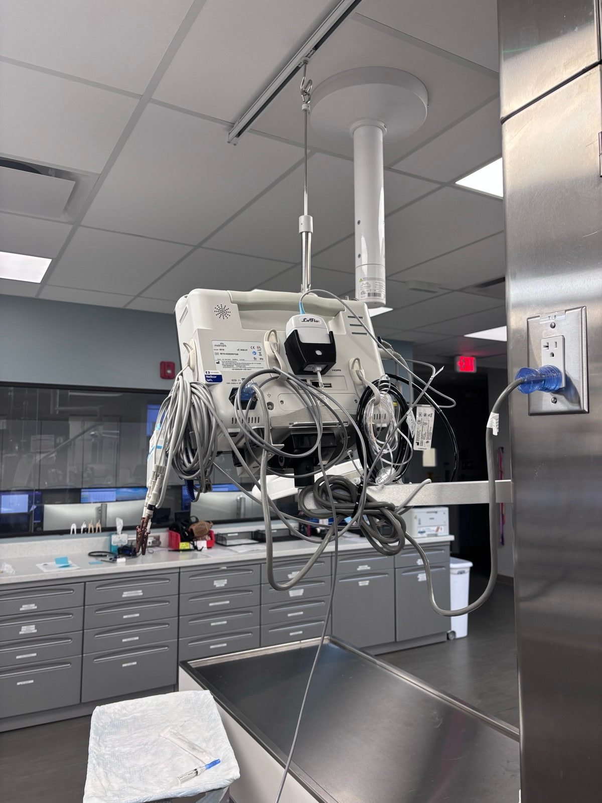
The Dangling Sensor Problem
The LoFlo module connects to compatible monitors through an 8-pin Lemo connector. It’s a precision instrument with infrared optics, a vacuum sampling pump, and sensitive electronics. And it ships with zero mounting hardware for any specific monitor.
In the real world, these modules end up sitting loose on top of the monitor, dangling from their cables, getting tucked behind equipment, or held in place with tape and zip ties. Every one of those situations causes problems.
A dangling module puts constant strain on the Lemo connector, causing intermittent electrical contact. Jostling creates vacuum leaks at the sampling line connection, which means false readings. And a module that gets knocked off and hits the floor can damage the infrared optics.
These aren’t edge cases. The Health Canada alert exists because cable damage from inadequate support is a real and documented failure mode.
Mounting the Sensor Properly
A CO2 sensor mount bolts to existing screw points on the rear panel of Midmark 8019 series monitors. It holds the LoFlo module in a fixed position, routes the cable cleanly, and takes the mechanical stress off the Lemo connector.
No drilling, no adhesive, no modification to the monitor. It also fits the Respironics CAPNOSTAT 5, plus most sidestream modules with a standard LoFlo-compatible form factor. Works with Bionet, Edan, and Infinium monitoring platforms as well.
For more on equipment organization issues like this, see 7 common clinic equipment problems.
Reading the Waveform: Four Patterns to Know
The capnogram waveform tells you more than the ETCO2 number alone. Four patterns worth memorizing:
Normal: A square-shaped wave. Flat baseline near zero (inspiration), sharp upstroke (early expiration), level plateau, sharp downstroke as the next breath starts. ETCO2 is the value at the end of the plateau. This is what you want to see.
Curare cleft: A small dip or notch in the plateau. The patient is trying to breathe on their own as neuromuscular blockade wears off. Not an emergency, but a signal that your paralytic is running out.
Shark fin: The upstroke is sloped and rounded instead of sharp. This means airway obstruction: bronchospasm, mucus plug, or a kinked endotracheal tube. The more gradual the slope, the worse the obstruction. Investigate immediately.
Rising baseline: Both the baseline and peak ETCO2 climb over successive breaths. Classic sign of exhausted soda lime in a circle breathing system. The absorbent isn’t removing CO2 from inspired gas anymore, so the patient is rebreathing. Replace it.
Where Capnography Fits in Your Monitoring Setup
The 2020 AAHA Anesthesia and Monitoring Guidelines recommend continuous ventilation monitoring for all anesthetized patients. Capnography is the most effective way to do that. It belongs alongside pulse oximetry, ECG, non-invasive blood pressure, and continuous temperature monitoring.
For the temperature monitoring side of things, see our post on post-operative hypothermia and the Bair Hugger cage door adapter for recovery warming. Browse monitoring accessories in our product catalog.
This article is for informational purposes only. VetBog products are accessories, not FDA-cleared medical devices. Always follow your facility’s clinical protocols and the 2020 AAHA Anesthesia and Monitoring Guidelines. Brand names are trademarks of their respective owners, used for equipment identification under nominative fair use.
Frequently Asked Questions
What is a normal ETCO2 for dogs under anesthesia?
35 to 45 mmHg. Above 60 mmHg means the patient needs help ventilating (manual breaths or lighter anesthetic plane). Below 20 mmHg is concerning for low cardiac output or cardiac arrest. Cats run a bit lower, typically 28 to 35 mmHg.
Can I add capnography to my existing Midmark monitor?
Yes, if your Midmark supports the LoFlo sidestream module (most 8019 series monitors do). The LoFlo plugs into the 8-pin Lemo connector on the monitor’s CO2 input. A sensor mount secures the module to the monitor housing using existing screw points.
Why does my CO2 reading drift during long procedures?
Check the sampling line first. Water accumulation and partial occlusion cause most drift issues. Other possibilities: sensor calibration drift, or an unsecured module with intermittent vacuum leaks at the connector.
Is sidestream or mainstream better for small animal veterinary?
Sidestream is generally preferred. The lightweight sampling line adds less dead space and puts less traction on the ET tube, which matters for cats, toy breeds, and small exotics. Sidestream modules can also measure anesthetic gases alongside CO2.