Veterinary FAST Scan Workflow: How Equipment Disorganization Costs Critical Minutes
How disorganized FAST scan supplies slow veterinary ER triage. Protocols for AFAST, TFAST, and Vet BLUE, plus how to keep everything on the ultrasound unit itself.
What FAST Scans Are and Why They Matter
FAST (Focused Assessment with Sonography for Trauma) is a set of point-of-care ultrasound protocols developed by Dr. Gregory Lisciandro for veterinary emergency medicine. These aren’t comprehensive abdominal ultrasounds. They’re rapid, targeted assessments designed to answer specific yes-or-no questions at the bedside: Is there free fluid? Pericardial effusion? Pleural effusion? Are the lungs wet?
Three protocols make up the system:
- AFAST (Abdominal FAST): four-point abdominal scan for free fluid
- TFAST (Thoracic FAST): thoracic assessment for pericardial effusion, pleural effusion, and pneumothorax
- Vet BLUE (Veterinary Bedside Lung Ultrasound Exam): bilateral lung scanning for B-lines, consolidation, and other pathology
Used together, they’re called Global FAST. You’ll see them in trauma triage, acute collapse, hemorrhagic shock, respiratory distress, and post-surgical monitoring. These are the sickest patients in your ER.
The Three Protocols, Briefly
AFAST scans four quadrants with the patient in right lateral recumbency:
- DH (Diaphragmatic-Hepatic): subxiphoid, looking at the liver and diaphragm
- SR (Spleno-Renal): left flank, spleen and left kidney
- CC (Cysto-Colic): caudal midline, bladder and colon
- HR (Hepato-Renal): right flank, right kidney and liver
Each site is scored for free fluid on a 0 to 4 scale using the Abdominal Fluid Scoring system. A positive finding at any site triggers the next decision: abdominocentesis for fluid characterization, surgical consult, or serial monitoring.
TFAST looks at the thorax for pericardial effusion, pleural effusion, and pneumothorax. Vet BLUE scans four bilateral lung regions (caudodorsal, perihilar, middle, cranial) looking for B-lines. Three or more B-lines at a single site is significant for alveolar-interstitial fluid.
When Minutes Actually Change Things
A complete AFAST takes about two minutes in trained hands. A full Global FAST can be done in under six. The scan itself is quick. The part that isn’t quick is getting everything ready.
Think about when these scans happen. A dog comes in with acute collapse and pale gums. Possible splenic mass rupture, possible hemoabdomen. A cat in respiratory distress that could be pleural effusion, pneumothorax, or cardiogenic pulmonary edema. A post-op patient whose heart rate is climbing and pressure is falling.
In every one of these cases, the FAST scan result changes what you do next. Free fluid on AFAST might mean emergency surgery. Pericardial effusion on TFAST might mean pericardiocentesis. The scan gives you answers. But the time between “this patient needs a FAST scan” and “we have results” isn’t just scan time. It includes setup, supply gathering, and all the small delays that add up when the ultrasound cart isn’t ready.
Where the Supplies Actually Are
The scan itself needs a probe, coupling gel, and a clipped scan window. Simple enough. But the clinical workflow that follows the scan needs more. If AFAST shows free fluid, you want to tap it immediately: syringes for abdominocentesis (3 mL for diagnostic, 12 mL for therapeutic), hypodermic needles (22-gauge for sampling, 18-gauge for drainage), EDTA and red-top sample tubes, gauze, alcohol pads.
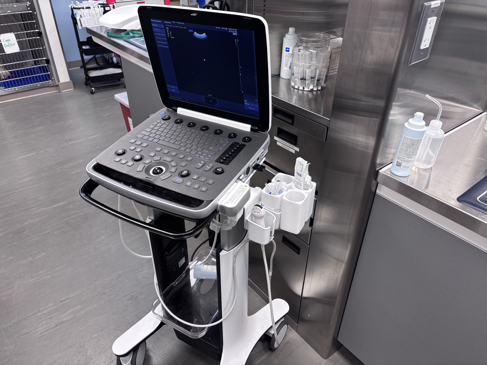
Portable veterinary ultrasound units come with minimal storage. A probe slot, maybe a gel holder. That’s it. Syringes are in a supply drawer across the room. Needles are in a cabinet that someone reorganized last Tuesday. Sample tubes are wherever the last person left them.
So the technician has to walk away from the patient to gather supplies. In a stable patient, that’s annoying. In a patient with hemorrhagic shock or cardiac tamponade, it’s a delay that matters.
The Repeat Scan Problem
FAST scans aren’t one-and-done. Post-surgical patients and critical care cases get serial monitoring: repeat scans to track whether free fluid is getting better, getting worse, or staying the same. A patient recovering from splenic surgery might get repeat AFAST throughout the first 12 to 24 hours.
Every repeat scan means restocking supplies. If the ultrasound machine moves between patients or rooms, supplies get left behind. If different technicians use the machine across shifts, nobody’s sure where things are.
You can train everyone to gather supplies before scanning. You can’t train a portable cart to have storage it was never designed with. This is a systems problem.
Going from Scan to Tap Without Leaving the Patient
The most important transition in a FAST scan workflow is the moment you go from imaging to intervention. AFAST reveals free fluid, and now you need to aspirate a sample. Is it blood? Transudate? Bile? Urine? The answer determines the entire treatment path.
If the supplies for abdominocentesis are already on the ultrasound unit (syringe, needle, sample tube, gauze), the technician transitions from scan to tap without ever leaving the patient’s side. If the supplies are in a drawer across the room, there’s a break in contact with a patient who may be actively decompensating.
A FAST scan pocket extension holds probes, coupling gel, syringes, needles, sample tubes, and gauze directly on the portable ultrasound unit. Everything goes where the machine goes.
What Should Be on Your Ultrasound Cart
Whether you use a dedicated organizer or not, here’s what should live on or right next to your portable ultrasound for FAST scans:
For the scan:
- Microconvex or curvilinear probe
- Coupling gel
- Clippers (for scan windows)
- Alcohol prep pads
For abdominocentesis:
- 3 mL syringes (diagnostic)
- 12 mL syringes (therapeutic)
- 22-gauge needles, 1 to 1.5 inch (diagnostic)
- 18-gauge needles (therapeutic, larger volumes)
- EDTA tubes (cytology)
- Red-top/no-additive tubes (culture)
- Gauze pads
If you might need it during or immediately after a FAST scan, it belongs on the machine. Not in a drawer. Not across the room. For related monitoring equipment topics, see our capnography guide and the CO2 sensor mount for Midmark patient monitors.
Making It Stick Across Your Team
Organization only works if everyone does it the same way. Three things to standardize:
- Supply locations: same supplies, same spots, every shift. When someone reaches for a syringe in a high-stress emergency, muscle memory matters.
- Protocol card: a laminated AFAST/TFAST/Vet BLUE reference card attached to the machine. Newer staff shouldn’t have to memorize scan points under pressure.
- Post-scan restocking: whoever finishes a scan replaces any used supplies before parking the machine. No exceptions.
Image acquisition for FAST scans can be performed by trained veterinary technicians. Interpretation and clinical decision-making remain the veterinarian’s responsibility.
The Simple Version
The ultrasound unit is the mobile platform. It goes to the patient. The supplies should go with it.
For a broader look at workflow problems like this, see 7 common veterinary clinic equipment problems. Browse all accessories at our product catalog.
This article is for informational purposes only. VetBog products are accessories, not FDA-cleared medical devices. Always follow your facility’s clinical protocols. Brand names are trademarks of their respective owners, used for equipment identification under nominative fair use.
Frequently Asked Questions
What is the difference between AFAST and TFAST?
AFAST scans four abdominal quadrants looking for free fluid. TFAST looks at the thorax for pericardial effusion, pleural effusion, and pneumothorax. Both use the same curvilinear or microconvex probe and are usually performed together as part of a Global FAST exam.
How often should post-surgical patients get repeat FAST scans?
There’s no universal interval. It depends on the patient. Repeat scans are indicated whenever a patient’s status changes, or to track whether free fluid is progressing or resolving. Daily scans are common after abdominal surgery, with more frequent checks for unstable patients.
Can veterinary technicians perform FAST scans?
Technicians can be trained to acquire the images (the scanning itself). Interpretation and clinical decisions based on the findings are the veterinarian’s responsibility. This division is standard in most veterinary ER settings.
What supplies should be stocked on a portable ultrasound unit?
At minimum: coupling gel, 3 mL and 12 mL syringes, 22-gauge and 18-gauge needles, EDTA and red-top sample tubes, gauze, and alcohol pads. Clippers should be within reach too. A FAST scan pocket extension keeps all of this organized on the machine itself.